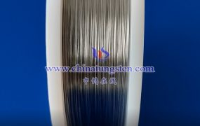
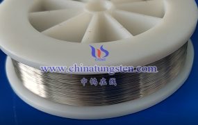
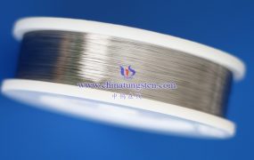
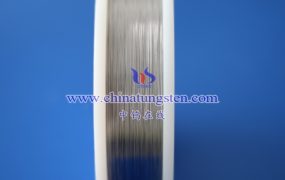
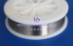
The microstructure analysis of tungsten wire surface usually involves multiple steps and multiple analysis techniques. The following is a detailed description of the analysis method of tungsten wire surface microstructure:
- Sample preparation
Sampling: Cut a small section from the tungsten wire to be tested as a sample to ensure that the sample is representative.
Cleaning: Use appropriate cleaning agents (such as alcohol, acetone, etc.) to clean the sample to remove oil and impurities on the surface.
Drying: Dry the cleaned sample to avoid the influence of moisture on subsequent analysis.
- Analysis method
Optical microscope observation:
Use an optical microscope to make a preliminary observation of the tungsten wire sample, and you can observe the morphology of the material surface, the preliminary distribution of grains, and possible defects.
The magnification of the optical microscope is moderate, which is suitable for preliminary transition observation of the tungsten wire surface from macro to micro.
Scanning electron microscope (SEM) observation:
Scanning electron microscope has higher magnification and resolution, and can observe more subtle structures on the surface of tungsten wire.
Through SEM observation, the grain morphology, hole distribution, and possible defects such as cracks and oxide layers on the surface of tungsten wire can be clearly seen.
SEM can also be combined with energy dispersive spectrometer (EDS) for elemental composition analysis to further understand the chemical composition of the surface of tungsten wire.
Transmission electron microscope (TEM) observation:
Transmission electron microscope has higher resolution and can observe the microstructure inside tungsten wire, such as lattice arrangement, dislocation, etc.
TEM is a powerful tool for occasions where in-depth understanding of the internal structure of tungsten wire is required.
However, it should be noted that the preparation process of TEM samples is relatively complicated, and thin slices need to be cut and thinned.
X-ray diffraction (XRD) analysis:
X-ray diffraction analysis can determine the crystal structure and phase composition of tungsten wire.
Through the XRD spectrum, information such as the lattice constant and interplanar spacing of tungsten wire can be obtained, and its crystal structure and phase composition can be inferred.
XRD analysis can also be used to detect possible impurity phases or second phases in tungsten wire.
Other analysis methods:
Atomic force microscopy (AFM), laser confocal microscopy, etc. can also be used for microstructure analysis of tungsten wire surface, but the specific method to be selected needs to be determined according to the analysis requirements and experimental conditions.
- Analysis results and discussion
Grain morphology and distribution:
By observing and analyzing the grain morphology and distribution on the surface of tungsten wire, we can understand the influence of its preparation process and heat treatment process on the grain morphology.
The size, shape and distribution of grains have an important influence on the properties of tungsten wire (such as strength, toughness, conductivity, etc.).
Defect analysis:
By observing and analyzing defects such as cracks and holes on the surface of tungsten wire, we can understand the possible problems in its preparation and processing process.
The presence of defects will affect the performance and service life of tungsten wire, so defects need to be strictly controlled.
Element composition analysis:
The chemical composition and impurity content of the tungsten wire surface can be understood by combining SEM with EDS or XRD and other methods.
Changes in elemental composition will affect the performance and stability of tungsten wires, so the elemental composition needs to be strictly controlled.
More details of tungsten wires, please visit website: http://tungsten.com.cn/tungsten-wires.html
Please contact CHINATUNGSTEN for inquiry and order of tungsten needles:
Email: sales@chinatungsten.com
Tel.: +86 592 5129595
